Scientists peek inside the mind of Maxwell’s demon

Physicists have now probed the memory of Maxwell’s demon, a devious, hypothetical beast. By peeking at information retained by a laboratory version of the creature, scientists confirmed the role of information in saving the second law of thermodynamics from the onslaught of a tiny, superpowerful being intent on wreaking havoc.

In work reported online July 3 in the Proceedings of the National Academy of Sciences, a team of researchers created a quantum version of Maxwell’s demon in the lab and measured both the information stored in its memory and the energy it extracted from a system. The results directly illustrate that information plays a key role in the demon’s attempts to distill energy.
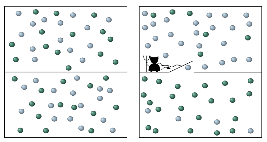
Since 1867, when the demon was proposed by physicist James Clerk Maxwell, scientists wondered whether such a creature could violate the second law, a sacred tenet of physics. It declares that the entropy, or disorder, of a closed system cannot decrease over time.

Maxwell suggested that a nefarious tiny being could shuttle around molecules to decrease entropy — for example, by putting all the fast-moving molecules on one side of a box containing a gas and the slower ones on the other side. Such an improbable reconfiguration would break the second law, allowing the demon to illegally siphon off energy.

A century later, a solution to this dilemma was found: The demon must record information about the molecules in order to manipulate them, and that information has physical relevance. Storing that information in its “brain” increases the entropy of the demon, compensating for the entropy decrease the demon produces. As the demon extracts energy, it must delete the contents of its memory in order to store new information and manipulate other molecules. That deletion, physicist Rolf Landauer determined in 1961, costs energy and releases entropy, with the result that the demon’s energy harvest is negated.

To show that the demon indeed remembered the properties of the system, the researchers probed the quantum state of the demon’s memory.

“The state of the memory is very important,” says physicist Juan Parrondo of Complutense University of Madrid, because it is what confirms that the second law still holds. “This is the first experiment which really addresses this question,” says Parrondo, who was not involved with the research.
In the experiment, performed by physicist Benjamin Huard and colleagues, the demon extracts energy from the system, a tiny circuit made of superconducting metal, which can carry electricity without resistance. Light tuned to a particular frequency causes the system to jump from a low-energy to high-energy state, or vice versa, absorbing or emitting a photon, or particle of light, in the process. The demon — a superconducting cavity within which microwaves bounce back and forth — manipulates the system to ensure that energy can be drained from the system, but not absorbed, allowing the demon to capture the energy released.

If the system is in the high-energy state, the demon allows the system to drop to lower energy, in the process spitting out a photon, which the demon can harvest for energy. But if the system is in the low-energy state, the demon prevents it from absorbing photons. The net result: Energy is sapped from the circuit. But, says Huard, of École Normale Supérieure de Lyon in France, “the information the demon learns about the system is encoded into its memory.”

The researchers probed this memory through a process called quantum tomography — meaning that they repeated the experiment many times and cataloged the state of the memory. The results revealed that, as expected, the demon retained the information about what energy state the system was in.

In addition to reading the demon’s mind, the researchers measured the work extracted from the system in a more direct manner than previous experiments. “This is one new feature of this implementation,” says Roberto Serra of Federal University of ABC in Santo André, Sao Paulo, Brazil. “This work is a very nice experiment.”

In Huard’s scheme — in contrast to some previous experiments — the demon and the system both operate on a quantum level. While the rules of thermodynamics were originally understood only for large systems like steam engines, scientists now hope understanding how the rules translate to small scales could one day lead to designs for more efficient quantum machines (SN: 3/19/16, p. 18).

Most football players who donated their brains to science had traumatic injury

A majority of football players whose brains were donated for research suffered a degenerative brain disease during their lives, according to the largest sample of players ever studied. The finding provides more evidence that the repetitive injuries to the brain sustained while playing American football are associated with the disease, researchers say.

Of 202 deceased former football players, 177 were diagnosed with chronic traumatic encephalopathy, which can cause a host of mood and behavioral issues as well as thinking and reasoning problems. Among 111 men who had played in the National Football League, 110 — a whopping 99 percent — had developed the disease, researchers report July 25 in JAMA. Three of 14 high school players also showed signs of the brain disease, as well as 48 of 53 college players. Researchers relied on brain autopsies of the players to make the diagnoses and interviewed family members and friends about the symptoms players had experienced.
This doesn’t necessarily mean all football players experience chronic traumatic encephalopathy. Many of the families who donated the brains for research could have been motivated to do so because their loved ones had noticeable symptoms, so the sample is not necessarily representative of the general football population. The results are still worrisome, though, researchers say.
“The fact that chronic traumatic encephalopathy was so common adds to our concern about the safety of playing football and the risk of developing neurologic symptoms later in life,” says neurologist Gil Rabinovici of the University of California, San Francisco, who wrote an editorial accompanying the article. This “hovers like a dark cloud over the game at all levels, even if the study cannot address how frequent the disease is, or who is at risk.”

Chronic traumatic encephalopathy, or CTE, shows up in athletes and others who’ve had repetitive injuries to the head, such as concussions. The only way to diagnose the disease is with an autopsy. In brains with the condition, a protein called tau goes “bad” and forms clumps in nerve cells and other brain cells. Although tau buildup is found in other brain diseases, like Alzheimer’s, in CTE, the protein congregates in brain cells around small blood vessels.
In 2008, a research team set up a brain bank to study the impact of head blows resulting from contact sports or military service. Behavioral neurologist Jesse Mez of Boston University School of Medicine and his colleagues classified players as having mild or severe CTE, depending on how widespread the tau clumps were in the players’ brains. The severity of disease seemed to track with the number of years spent playing football, says Mez. Among NFL players, 95 of the 110 diagnosed cases were severe. All three of the high school players’ cases were mild, while just over half of the college players’ cases were severe.

Yet the players’ reported symptoms while alive were similar, regardless of the severity seen in the brain. Behavioral and mood problems, such as impulsivity, anxiety and depression, were commonly reported in both severe and mild cases of the disease. Cognitive symptoms, including memory loss, were also typical for both groups. One major difference, Mez notes, was that dementia was more common in severe cases of CTE than in mild cases.

As for why players reportedly experienced similar symptoms no matter the severity, “the question is, is there something else going on,” such as inflammation, Mez says. “Or are there regions of the brain that we’re not looking carefully enough at?”

There still isn’t a way to diagnose CTE during life, and that’s “the 800-pound gorilla in the room,” says neurologist David Brody of Washington University School of Medicine in St. Louis.

Detecting the disease in patients will be crucial for understanding how common CTE is in the NFL, “let alone in the millions of people who participated in college, high school and youth football,” says Rabinovici. “In the meantime, we need to focus on prevention of concussions and other head impacts at all levels of contact sports.”

Fossil find suggests this ancient reptile lurked on land, not in the water

A round belly, stubby feet and a tapering tail made one armored reptile a lousy swimmer. Despite earlier reports, Eusaurosphargis dalsassoi might not have swum at all, scientists now say.

E. dalsassoi was first identified in 2003. Fossils were found near Monte San Giorgio at the Swiss-Italian border alongside the remains of marine reptiles and fish that lived roughly 240 million years ago. That association led scientists to conclude the creature was aquatic. But a complete skeleton of E. dalsassoi unearthed in 2002 in the Swiss Alps and recently assembled contradicts that idea.
At just under 20 centimeters long, the fossil, probably of a youngster, shows that E. dalsassoi widened at the stomach and slithered forward with stiff elbow and knee joints and spadelike claws. That’s not a swimmer’s build, paleontologist Torsten Scheyer of the University of Zurich and colleagues report June 30 in Scientific Reports.

Armed with rows of small spikes along its back and spear-shaped plates framing its head, sides and tail, the animal resembled today’s girdled lizards. The researchers speculate that this particular E. dalsassoi died on a beach and then got washed into the ocean.

How an itch hitches a ride to the brain

Scientists have traced the sensation of itch to a place you can’t scratch.

The discomfort of a mosquito bite or an allergic reaction activates itch-sensitive nerve cells in the spinal cord. Those neurons talk to a structure near the base of the brain called the parabrachial nucleus, researchers report in the Aug. 18 Science. It’s a region that’s known to receive information about other sensations, such as pain and taste.

The discovery gets researchers one step closer to finding out where itch signals ultimately end up. “The parabrachial nucleus is just the first relay center for [itch signals] going into the brain,” says study coauthor Yan-Gang Sun, a neuroscientist at the Chinese Academy of Sciences in Shanghai.
Understanding the way these signals are processed by the brain could someday provide relief for people with chronic itch, Sun says. While the temporary itchiness of a bug bite is annoying, longer term, “uncontrollable scratching behavior can cause serious skin damage.”

Previous studies have looked at the way an itch registers on the skin or how neurons convey those sensations to the spinal cord. But how those signals travel to the brain has been a trickier question, and this research is a “major step” toward answering it, says Zhou-Feng Chen, director of the Center for the Study of Itch at Washington University School of Medicine in St. Louis.

A network of neurons in the spinal cord wrangles itch signals, previous research suggests. In particular, spinal neurons that make a protein called gastrin-releasing peptide receptor have been shown to be important in itch signaling. But those neurons didn’t link up directly to the parabrachial nucleus, or PBN, Sun’s team found; instead, they talked to other neurons that send messages to the PBN.

When mice were given injections of a drug that induces allergic itching, the rodents showed greater activity in those neurons connecting the spinal cord to the PBN, Sun and colleagues found. In another experiment, the researchers made neurons going to the PBN light-sensitive, and then used light to stop those neurons from sending messages. When those nerve cells were blocked, mice given an itch-triggering drug scratched less.

It’s too soon to say whether itch signals in humans follow the same route — or whether all kinds of itches take the same path. An allergic itch is different from the sort of itch that comes from a light touch, and the two might be handled differently by the brain (SN: 11/22/08, p. 16). And mice, unlike humans, can’t actually describe how itchy they’re feeling. So scientists have to rely on clues like scratching, a reaction to an itch, not a direct measurement of the sensation itself. Which raises the question: If you don’t feel an urge to scratch an itch, is the itch really there at all?

Here’s what the Science News family did for the eclipse

We came. We saw. We earned our 2017 eclipse t-shirts.

For us, the Aug. 21 total solar eclipse was the culmination of weeks — nay, months — of planning the stories you’ve recently seen on Science News. And as the big day finally approached, many members of the Science News staff past and present traveled far and wide to experience the spectacle.

Undeterred by traffic and clouds, and fueled by bottomless fried chicken and moon pies, correspondents made their way to small towns and big cities in the path of totality. For many of us, this was our first total solar eclipse, and it lived up to the hype. Astronomy writer Lisa Grossman wrote from her perch on a mountaintop in Wyoming, “I thought I knew what to expect from my first total solar eclipse. I had no idea.”

Even the crew at the Science News offices in Washington, D.C., got a pretty sweet view of the moon blocking out 81 percent of the sun. And it has inspired many of us to start planning now for the total solar eclipse that will cross the United States in April 2024.

All in all, the 2017 eclipse was unlike anything we’ve seen before, and it’s something we’re not likely to forget. But just in case, the video below showcases some snapshots from our trips to totality and back again.

Rising temperatures threaten heat-tolerant aardvarks

When nocturnal aardvarks start sunbathing, something’s wrong.

If the animals are desperate enough to bask like some cold, sluggish turtle, it’s because they’ve got the chills. Robyn Hetem, an ecophysiologist, has the body temperature data to prove it — collected from late 2012 into 2013, the hottest summer the arid Kalahari region in South Africa had seen in more than 30 years.

Hotter, drier conditions are predicted to become the norm for southern Africa as the climate changes. Now Hetem and colleagues have used that foretaste of change to show that higher temperatures might hammer the normally heat-tolerant aardvarks by shrinking the animals’ food supply.
Aardvarks live their burrow-digging lives just about anywhere in sub-Saharan Africa except the desert. The toothless night-foragers dine by slurping insect colonies. One of Hetem’s students at the University of the Witwatersrand in Johannesburg spent two years collecting hundreds of aardvark droppings and can confirm that Orycteropus afer in the Kalahari eat only termites and ants. Yet the solitary, long-snouted, knee-high mammals are more closely related to elephants than to any pointy-nosed South American anteater.

An aardvark looks “very lethargic but is incredibly strong and fast,” Hetem says. The researchers wanted to fit wild aardvarks with tracking devices and data loggers but first had to catch the animals. Nets failed. Traps failed. One cornered aardvark burst out of a burrow, knocked four men to the ground and then outran them.
Eventually, researchers placed instruments on six animals. When the Kalahari baked and good rains were months late, the aardvarks grew thin and bony. They started hunting during the day and sunbathing. The animals, once able to internally stabilize their body temperatures, started to have great plunging chills at night, according to data loggers. That’s a sign of starvation, Hetem says, and occurs when the body no longer has energy to warm itself. Five of the six tracked animals died, along with at least 11 other aardvarks in the neighborhood.
Aardvark heat tolerance wasn’t the problem. The animals were dying off because their food couldn’t take the heat and drought, Hetem and colleagues argue in the July Biology Letters. Hot, dry spells can make ant and termite colonies shrink and retreat to hard-to-reach hideouts.
Other African wildlife might suffer from a shortage of aardvarks, which are prodigious burrow diggers. In a Kalahari study, one aardvark used more than 100 burrows in two years. So many hideaways are a boon for others. Bat-eared foxes, warthogs, birds called ant-eating chats and at least two dozen other species pop into aardvark architecture, sometimes outright moving in. If aardvarks dwindle, shelter might grow scarcer for other animals.

“We kind of think of climate change as: Things are going to get hotter and species might be sensitive to it,” Hetem says. “There’s so much more we need to understand.”

A researcher reveals the shocking truth about electric eels

Kenneth Catania knows just how much it hurts to be zapped by an electric eel. For the first time, the biologist at Vanderbilt University in Nashville has measured the strength of a defensive electrical attack on a real-life potential predator — himself.

Catania placed his arm in a tank with a 40-centimeter-long electric eel (relatively small as eels go) and determined, in amperes, the electrical current that flowed into him when the eel struck. At its peak, the current reached 40 to 50 milliamperes in his arm, he reports online September 14 in Current Biology. This zap was painful enough to cause him to jerk his hand from the tank during each trial. “If you’ve ever been on a farm and touched an electric fence, it’s pretty similar to that,” he says.
This is Catania’s latest study in a body of research analyzing the intricacies of an electric eel’s behavior. The way electric eels have been described by biologists in the past has been fairly primitive, says Jason Gallant, a biologist who heads the Michigan State University Electric Fish Lab in East Lansing who was not involved in the study. Catania’s work reveals that “what the electric eel is doing is taking the electric ability that it has and using that to its absolute advantage in a very sophisticated, deliberate way,” he says.

Electric eels use electric current to navigate, communicate and hunt for small prey. But when faced with a large land-based predator, eels will launch themselves from the water and electrify the animal with a touch of the head.
Using electrical measurements he collected during the eel attacks, Catania came up with an equation to estimate the amount of electric current flowing from the eel into his arm. The electric shock was strongest when the electric eel was farthest out of the water. That makes sense because when an eel is mostly submerged, the majority of the electricity dissipates in the water. As the eel rises out of the water, the only place left for the electricity to flow is into whatever the fish head-bumps (SN Online: 6/9/16).

Catania cannot say, however, whether a leap attack from an electric eel is equally as shocking for all potential predators. Electrical currents travel through an animal more or less effectively depending on its outer layer. The internal resistance, or opposition to electrical current flow, may be different for a human arm than for an animal with scales or fur, like a crocodile or a dog, Catania notes. More research is needed to understand how powerful the shock is for other land animals.
Extrapolating from his experience with a small eel, Catania estimates that a human struck on the trunk by a larger, 1.8-meter-long electric eel might endure a current of 0.24 amperes, or 63 watts of power. That’s about 8½ times as powerful as the zap from a typical law-enforcement Taser gun.

Gene variant linked to Alzheimer’s disease is a triple threat

A genetic risk factor for Alzheimer’s disease is a double, make that triple, whammy.

In addition to speeding up the development of brain plaques associated with Alzheimer’s, a gene variant known as APOE4 also makes tau tangles — another signature of the disease — worse, researchers report online September 20 in Nature. APOE4 protein also ramps up brain inflammation that kills brain cells, neuroscientist David Holtzman of Washington University School of Medicine in St. Louis and colleagues have discovered.
“This paper is a tour de force,” says Robert Vassar, a neuroscientist at Northwestern University Feinberg School of Medicine in Chicago. “It’s a seminal study that’s going to be a landmark in the field” of Alzheimer’s research, Vassar predicts.

For more than 20 years, researchers have known that people who carry the E4 version of the APOE gene are at increased risk of developing Alzheimer’s. A version of the gene called APOE3 has no effect on Alzheimer’s risk, whereas the APOE2 version protects against the disease. Molecular details for how APOE protein, which helps clear cholesterol from the body, affects brain cells are not understood.

But Holtzman and other researchers previously demonstrated that plaques of amyloid-beta protein build up faster in the brains of APOE4 carriers (SN: 7/30/11, p. 9). Having A-beta plaques isn’t enough to cause the disease, Holtzman says. Tangles of another protein called tau are also required. Once tau tangles accumulate, brain cells begin to die and people develop dementia. In a series of new experiments, Holtzman and colleagues now show, for the first time, that there’s also a link between APOE4 and tau tangles.
In one experiment, mice that had no A-beta in their brains developed more tau tangles if they carried the human version of APOE4 than if they had the human APOE3 gene, Holtzman and colleagues found. That finding indicates APOE4 affects tau independently of A-beta.
Brains of people who died from various diseases caused by tangled tau had more dead and damaged cells if the people carried APOE4. The researchers also tracked 592 people who had low levels of A-beta in their cerebral spinal fluid — a clue that plaques have formed in the brain — and who showed symptoms of Alzheimer’s. Over a five- to 10-year period, the disease progressed 14 percent faster in people with one copy of APOE4 and 23 percent faster in people with two copies than in people who didn’t have that version of the gene, the researchers found. Those worsening symptoms are presumed to be caused by more rapid buildup of tau tangles in the APOE4 carriers.

APOE4 also seems to make Alzheimer’s worse by causing inflammation, the researchers found. Two kinds of mouse glial brain cells, microglia and astrocytes, making different versions of the APOE protein were grown with brain nerve cells, or neurons, that make disease-causing forms of tau. Mouse neurons grown with glia making no APOE grew well, even though they were making abnormal tau. But neurons grown with glia making APOE4 often died. APOE4 provoked inflammation responses in the normally friendly astrocytes and microglia, leading those cells to kill neurons, the researchers found. Such inflammation can make brain degeneration worse.

The data linking the APOE4 gene to tau tangles and brain inflammation is “super tight,” says molecular neurobiologist Sangram Sisodia of the University of Chicago. But the molecular details behind how APOE4 protein causes those effects are still vexingly absent, he says. Much more work is needed to uncover which molecules APOE4 interacts with, so that researchers can devise ways to counteract its negative effects in the brain.

Any therapies that decrease or eliminate APOE4 will need to be limited to the brain, because the protein is needed in the rest of the body to maintain healthy cholesterol levels, Vassar says. “You don’t want to give a person heart disease to cure Alzheimer’s disease.”

How a meteor shower helped solve the case of the vanishing comet

The reappearance of a long-lost meteor shower has finally explained what happened to a missing comet named 289P/Blanpain.

That comet was spotted only once in 1819 and never again, unusual for a body orbiting the sun. But in 2003, astronomers found a small asteroid moving along the Blanpain orbit, suggesting the space rock might be the comet (or a piece of it) after it ejected much of its cometary dust.

Some of that dust may have been what Japanese researchers saw in 1956 when they observed a meteor shower from the constellation Phoenix. Meteor showers occur when dust left behind by a comet burns up as it hits Earth’s atmosphere. Those “Phoenicid” meteors hadn’t been seen before — or since.
Astronomer Jun-ichi Watanabe of the National Astronomical Observatory of Japan in Tokyo and colleagues traced the meteors to where the comet’s dust trail should have been. In 2010, the group predicted that the remaining dust would create another shower in 2014.

Team members traveled to North Carolina and Spain’s Canary Islands to test their prediction, and on the first two days of December, 2014, they saw Phoenicids streak across the sky. But there were about 90 percent fewer meteors than expected; Blanpain may have lost its dust more quickly than previously thought, the team reports in the Sept. 1 Planetary and Space Science. The astronomers will get a second chance to check — another shower is expected in 2019.