A genetic risk factor for Alzheimer’s disease is a double, make that triple, whammy.
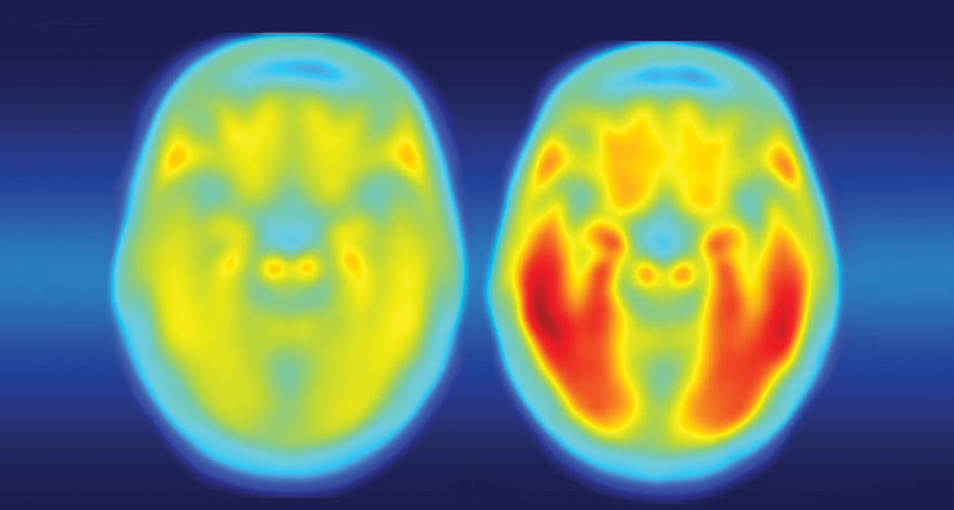
In addition to speeding up the development of brain plaques associated with Alzheimer’s, a gene variant known as APOE4 also makes tau tangles — another signature of the disease — worse, researchers report online September 20 in Nature. APOE4 protein also ramps up brain inflammation that kills brain cells, neuroscientist David Holtzman of Washington University School of Medicine in St. Louis and colleagues have discovered. “This paper is a tour de force,” says Robert Vassar, a neuroscientist at Northwestern University Feinberg School of Medicine in Chicago. “It’s a seminal study that’s going to be a landmark in the field” of Alzheimer’s research, Vassar predicts.
For more than 20 years, researchers have known that people who carry the E4 version of the APOE gene are at increased risk of developing Alzheimer’s. A version of the gene called APOE3 has no effect on Alzheimer’s risk, whereas the APOE2 version protects against the disease. Molecular details for how APOE protein, which helps clear cholesterol from the body, affects brain cells are not understood.
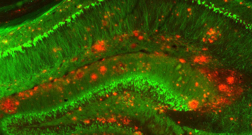
But Holtzman and other researchers previously demonstrated that plaques of amyloid-beta protein build up faster in the brains of APOE4 carriers (SN: 7/30/11, p. 9). Having A-beta plaques isn’t enough to cause the disease, Holtzman says. Tangles of another protein called tau are also required. Once tau tangles accumulate, brain cells begin to die and people develop dementia. In a series of new experiments, Holtzman and colleagues now show, for the first time, that there’s also a link between APOE4 and tau tangles. In one experiment, mice that had no A-beta in their brains developed more tau tangles if they carried the human version of APOE4 than if they had the human APOE3 gene, Holtzman and colleagues found. That finding indicates APOE4 affects tau independently of A-beta. Brains of people who died from various diseases caused by tangled tau had more dead and damaged cells if the people carried APOE4. The researchers also tracked 592 people who had low levels of A-beta in their cerebral spinal fluid — a clue that plaques have formed in the brain — and who showed symptoms of Alzheimer’s. Over a five- to 10-year period, the disease progressed 14 percent faster in people with one copy of APOE4 and 23 percent faster in people with two copies than in people who didn’t have that version of the gene, the researchers found. Those worsening symptoms are presumed to be caused by more rapid buildup of tau tangles in the APOE4 carriers.
APOE4 also seems to make Alzheimer’s worse by causing inflammation, the researchers found. Two kinds of mouse glial brain cells, microglia and astrocytes, making different versions of the APOE protein were grown with brain nerve cells, or neurons, that make disease-causing forms of tau. Mouse neurons grown with glia making no APOE grew well, even though they were making abnormal tau. But neurons grown with glia making APOE4 often died. APOE4 provoked inflammation responses in the normally friendly astrocytes and microglia, leading those cells to kill neurons, the researchers found. Such inflammation can make brain degeneration worse.
The data linking the APOE4 gene to tau tangles and brain inflammation is “super tight,” says molecular neurobiologist Sangram Sisodia of the University of Chicago. But the molecular details behind how APOE4 protein causes those effects are still vexingly absent, he says. Much more work is needed to uncover which molecules APOE4 interacts with, so that researchers can devise ways to counteract its negative effects in the brain.
Any therapies that decrease or eliminate APOE4 will need to be limited to the brain, because the protein is needed in the rest of the body to maintain healthy cholesterol levels, Vassar says. “You don’t want to give a person heart disease to cure Alzheimer’s disease.”
The reappearance of a long-lost meteor shower has finally explained what happened to a missing comet named 289P/Blanpain.
That comet was spotted only once in 1819 and never again, unusual for a body orbiting the sun. But in 2003, astronomers found a small asteroid moving along the Blanpain orbit, suggesting the space rock might be the comet (or a piece of it) after it ejected much of its cometary dust.
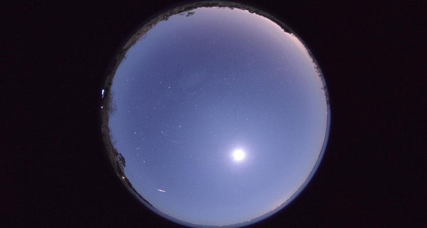
Some of that dust may have been what Japanese researchers saw in 1956 when they observed a meteor shower from the constellation Phoenix. Meteor showers occur when dust left behind by a comet burns up as it hits Earth’s atmosphere. Those “Phoenicid” meteors hadn’t been seen before — or since. Astronomer Jun-ichi Watanabe of the National Astronomical Observatory of Japan in Tokyo and colleagues traced the meteors to where the comet’s dust trail should have been. In 2010, the group predicted that the remaining dust would create another shower in 2014.
Team members traveled to North Carolina and Spain’s Canary Islands to test their prediction, and on the first two days of December, 2014, they saw Phoenicids streak across the sky. But there were about 90 percent fewer meteors than expected; Blanpain may have lost its dust more quickly than previously thought, the team reports in the Sept. 1 Planetary and Space Science. The astronomers will get a second chance to check — another shower is expected in 2019.
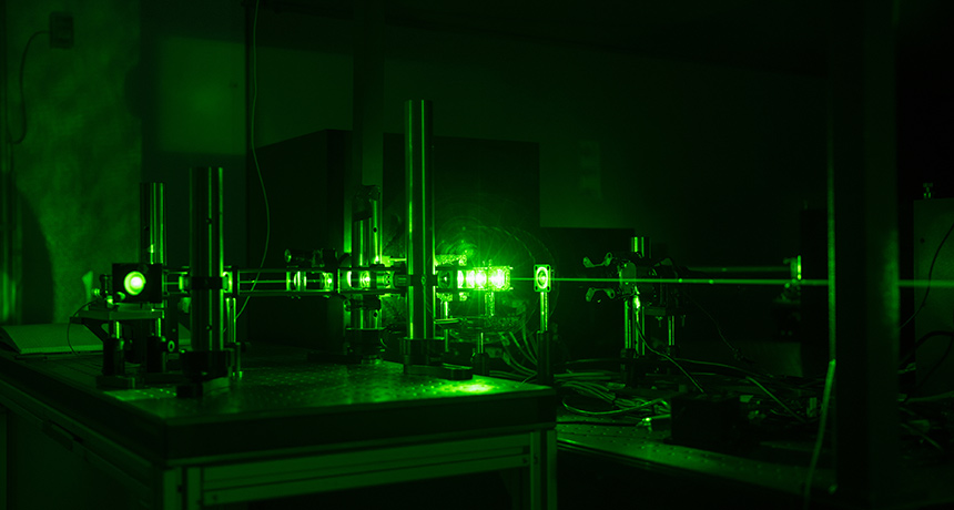
Light is two-faced: Sometimes it behaves like a wave, sometimes like a particle. Now, scientists have shown that light’s shifty disposition persists even after trekking thousands of kilometers into space and back again, researchers report October 25 in Science Advances.
Depending on how light is measured, it can either be particle-like, lighting up a camera pixel, for example, or wavelike, interfering with other waves like ripples on the surface of water. It’s one of the many oddities of quantum mechanics. Before light is measured, quantum theory suggests, it is in a particle-wave limbo, neither purely one nor the other.
Physicists have tested this idea by performing “delayed-choice” experiments in the lab, in which researchers send light into a device and randomly choose whether or not to flip a switch that seems to retroactively change the light’s behavior (SN: 5/30/15, p. 9). In one configuration, the light travels down two paths at once and acts like a wave, interfering with itself. In the other, the light acts like a particle, taking a single path. That choice of configuration can be made even after the light has already traveled through the device but before being measured, revealing that light remains in quantum limbo until it is finally detected.
For the first time, physicist Paolo Villoresi of the University of Padua in Italy and colleagues took the technique into space. The researchers sent light through a lab apparatus and up to a satellite equipped with reflectors, which bounced the light back down to the device. While the light was in transit, the scientists used a random number generator to determine whether to configure their apparatus so that the light would behave like a particle or a wave. The light performed as expected, verifying that quantum mechanics holds even over the round trip into space and back.
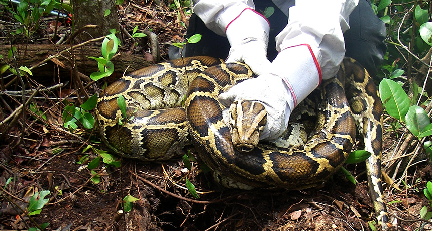
Remote Bouvet Island, a tiny, glacier-smothered landmass in the South Atlantic rimmed by 500-meter-tall cliffs, has a notable distinction: It’s the only known spot on Earth, scientists say, that has zero invasive species. Every other place, and every person, on the planet is at least indirectly affected by one or more species that has been transported — either intentionally or inadvertently — to new lands from the ecosystems in which the species evolved. In The Aliens Among Us, biologist and science journalist Leslie Anthony chronicles the detrimental effects of invasive species, as well as how these organisms spread and how they can be fought. In the United States, such interlopers — everything from zebra mussels in the Great Lakes to Burmese pythons in the Everglades — damage crops, infrastructure or otherwise cost taxpayers about $145 billion annually.
Invasive species, Anthony writes, are “children born of globalization and consumerism.” Their numbers increase as international trade widens and accelerates. Some species surreptitiously hitch a ride to their new homes on human transport: Think seeds and burrs on hikers’ clothing, or fish in ballast water of cargo ships. Others have been deliberately released, like earthworms or baitfish set loose by fishermen, or exotic lizards and snakes set free by careless pet owners. Rats, the world’s foremost invasive species, have traveled the world with explorers and traders; so have tropical fire ants, which genetic studies suggest have hitchhiked from southwestern Mexico to Asia and beyond starting in the 16th century in soil used as ballast in Spanish ships.
The Aliens Among Us is a thoroughly engaging book that draws from Anthony’s fieldwork and interviews with scientists, community volunteers, government researchers and policy makers. These groups are struggling to intercept species before they establish a beachhead on new shores, as well as eradicate those that have already gained a foothold. Discussion of people fighting the spread of Zika virus and other exotic diseases — big threats despite their minuscule size — makes the book especially timely. Some battles against invasives seem almost doomed to fail. Besides the inexorable increase of trade, the inescapable specter of climate change continues to open new vistas for species to colonize (SN: 12/24/16, p. 23).
An Alzheimer’s-related protein can move from the blood to the brain and accumulate there, experiments on mice show for the first time.
The results, published online October 31 in Molecular Psychiatry, suggest that the protein amyloid-beta outside the brain may contribute to the Alzheimer’s disease inside it, says Mathias Jucker, a neurobiologist at the University of Tübingen in Germany. This more expansive view of the disease may lead scientists to develop treatments that target parts of the body that are easier than the brain to access.
The experiments don’t suggest that people could contract Alzheimer’s from another person’s blood. “The bottom line is that this study is thought-provoking but shouldn’t cause alarm,” says neurologist John Collinge of University College London. “There really isn’t any evidence that you can transmit Alzheimer’s disease by blood transfusion.”
But researchers wondered whether, over time, A-beta might build up in the brain by moving there from the blood, where it’s normally found in small quantities. Earlier animal studies have shown that A-beta can move into the brain if it’s injected into the bloodstream, but scientists didn’t know whether A-beta from the blood can be plentiful enough to form plaques in the brain. To test this, researchers used a form of extreme blood-sharing in the experiment. Six pairs of mice — with one mouse engineered to produce gobs of human A-beta and one normal — were surgically joined for a year, causing blood mingling that’s far more extensive than that of a blood transfusion. After a year, the brains of the mice carrying the mutations were full of A-beta plaques, as expected. But these plaques were also inside the brains of the normal mice in the joined pairs. In those normal mice, A-beta levels weren’t as high as they were in the mutated mice, but the fact that they existed was notable, says study coauthor Weihong Song of the University of British Columbia in Vancouver. Unjoined control mice without the mutation showed no A-beta accumulation.
The brains of the joined mice also showed other signs of deterioration. The researchers observed inflammation, tiny areas of bleeding and a dangerous type of the protein tau in the brains of normal mice that had been exposed to blood full of A-beta. In people, Alzheimer’s is often marked by both A-beta plaques and tangles of tau. The results don’t mean that Alzheimer’s is predominantly caused by factors in the blood. “We still think of Alzheimer’s as a brain disorder,” Song says. But factors in the blood, in some cases, might have the power to nudge the disease along, the results suggest.
A-beta is made by cells in the brain, but also by blood platelets, skin cells, muscles and other parts of the body. Normally, “there is a balance between A-beta inside and outside the brain,” Song says. But when this balance is thrown off, such as when the body is chock-full of the protein, or when the blood-brain barrier — the blockade that keeps potential dangers out of the brain — deteriorates with age, the brain may get an extra dose, Song proposes. By tweaking this balance, it’s possible that drugs or therapies that reduce A-beta in the body might help slow or prevent Alzheimer’s disease.
Evidence has been accumulating that A-beta can behave as a prion, a misfolded protein that can incite normally folded proteins to go rogue (SN: 10/17/15, p. 12). Song says that the experiments don’t address whether A-beta from the blood can behave as a prion and prompt already existing A-beta in the brain to form plaques. The normal mice’s brain plaques seemed to be built from human A-beta protein, and the only source of that was the blood of the mutated partner mouse.
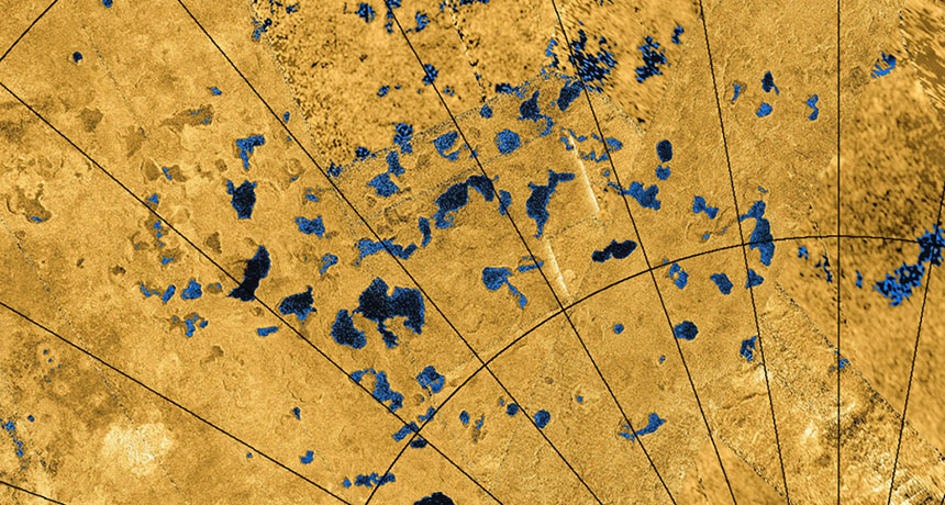
Liquid methane and ethane flow through a subterranean plumbing system on Titan, which drains lakes and connects seas. That’s one of the first scientific results from the latest, most complete map of the Saturnian moon’s topography.
Planetary scientist Paul Corlies of Cornell University and colleagues released the map — based on all the data from NASA’s Cassini mission, which ended in September (SN Online: 9/15/17) — in Geophysical Research Letters on December 2.
Titan, Saturn’s largest moon, hosts seas, lakes, clouds and rain — all composed of hydrocarbons such as methane and ethane instead of water. The elevations of seas and mountains across 9 percent of Titan’s surface were directly recorded by Cassini as it flew past Titan over 13 years. The researchers had to infer altitudes for the rest of the globe. Compared with previous maps, the new one adds mountains in the southern hemisphere and shows that Titan is more of a squashed sphere than previously thought. Researchers can now use the map to build computer simulations of everything from Titan’s atmosphere to its interior structure. “Within hours of the paper actually being available online, people we’ve never collaborated with started contacting [Corlies] to ask how to get the data,” says study coauthor Alexander Hayes, a planetary scientist also at Cornell.
But the first study to use the map, also published December 2 in Geophysical Research Letters, is research that Hayes has been working on for a decade. The work shows that Titan has a sea level as well as the hydrocarbon equivalent of groundwater — pores in subsurface rock are filled with liquid that can seep into and between the lakes and seas.
“Looking for actual evidence that the lakes could be communicating was a fundamental question from Cassini,” Hayes says. “This is the final paper that gives the best evidence that it exists.” His team analyzed the altitudes of Titan’s liquid bodies and found that the three largest seas — Ligeia Mare, Kraken Mare and Punga Mare— are all about the same elevation, just like Earth’s oceans. In other words, Titan has a sea level, Hayes says. To maintain that uniformity, the seas must be connected through channels that could be above or below ground. The moon’s poles are dotted with small lakes and depressions that are shaped like lakes but contain no liquid. Hayes and colleagues found that the liquid levels of the filled lakes are above sea level, so they are potentially isolated from the seas. If the lakes and seas were connected, the lakes’ liquid could drain into the seas, and the liquids’ surface heights would all match — or the lakes would be empty.
The floors of the dry lake beds are at a higher elevation still. Hayes thinks that may indicate that their liquid flowed into the filled polar lakes. Those hydrological connections probably occur underground because there do not appear to be enough connections on the surface. If scientists could dig deeper into a dry lake, he predicts, they would hit liquid at the level of the filled lakes’ surfaces.
A remaining mystery is how the small polar lakes formed. Both the dry and filled lakes have steep walls, flat floors and rims that rise above the surrounding ground — features that lakes on Earth tend not to have. “They look like you went around Titan’s polar region with a cookie cutter and cut out little shapes,” Hayes says.
His best guess is that the lakes are sinkholes (SN: 1/25/14, p. 14), which collapsed when the bedrock material was dissolved out from under them. If true, then Titan’s poles may be covered with a thick layer of a kind of solid that hydrocarbons can dissolve, like acetylene. But sinkholes shouldn’t have raised rims, so that theory doesn’t explain everything.
The researchers hope other investigators will have some new ideas. “We’re just saying, these are all the observations. Please tell us how they fit together,” Hayes says.
Planetary scientist Jani Radebaugh of Brigham Young University in Provo, Utah, thinks the odd lakes could be the remnants of icy volcanoes. Explosive eruptions could create the raised rims, and the depressions could be empty magma chambers that collapsed. “I think we should consider it,” she says.
But she agrees that Hayes’ groundwater theory makes sense. Seeing hydrological systems on Titan that are similar to Earth’s “is satisfying, and helps to validate that what we understand from the Earth should work on other bodies, regardless of what the liquid is made of,” she says.
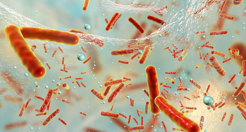
A new antibacterial ointment could help take down drug-resistant bacteria.
In human skin samples and mice, the medicine completely cleared wounds of MRSA, the strain of Staphylococcus aureus that is resistant to methicillin and other antibiotics, and antibiotic-resistant Acinetobacter baumannii. Both microbes are known to cause serious infections in hospital patients. Researchers in the Netherlands created the gel’s key ingredient, a chain of amino acids called SAAP-148, by improving on a bacteria-fighting peptide found in humans. The synthetic peptide prevents pathogens from forming biofilms — colonies of microbes enveloped in a protective slime that shields them from antibiotics, the researchers report online January 10 in Science Translational Medicine. Bacteria living in a biofilm can be 10 to 1,000 times as hard to kill as their free-floating counterparts. SAAP-148 also wiped out microbes that hunker down in a dormant, drug-tolerant state during an antibiotic assault, then lead the bacterial resurgence after treatment ends.
“This peptide could provide a much-needed boost to our arsenal of antibiotics,” says David Weiss, a microbiologist at Emory University School of Medicine in Atlanta, who wasn’t involved in the work.
Unlike S. aureus and A. baumannii bacteria exposed to conventional antibiotics in the study, the microbes didn’t develop strong resistance to SAAP-148 after at least a couple weeks’ exposure to the compound. These results are “quite promising,” says Weiss, who would like to see the peptide tested against other bacteria as well. Studies of the gel in humans will begin this year.
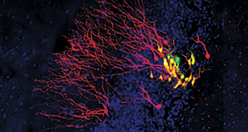
Brain scientists have filmed a first-of-a-kind birth video. It reveals specialized cells in the brains of mice dividing to create newborn nerve cells.
The images, published in the Feb. 9 Science, show intricacies of how certain parts of the adult mouse brain can churn out new nerve cells. These details may help lead to a deeper understanding of the role of this nerve cell renewal in such processes as memory.
Deep in the brains of mice, a memory-related structure called the hippocampus is known to be flush with new nerve cells. But because this buried neural real estate is hard to study, the circumstances of these births weren’t clear. Using living mice, Sebastian Jessberger, a neuroscientist at the University of Zurich, and colleagues removed the outer layers of brain tissue that obscure the hippocampus. The scientists marked 63 cells called radial stem cells, which can divide to create new nerve cells. Researchers then watched these stem cells for up to two months, taking pictures every 12 or 24 hours. During that time, 42 of these stem cells underwent a spurt of division, churning out two kinds of cells: intermediate cells that would go on to produce nerve cells as well as mature nerve cells themselves. Once this burst of activity ended, the radial stem cells disappeared by dividing themselves into mature nerve cells that could no longer split.
Many of these newly formed nerve cells had brief lives, dying either within the first four days, or 13 to 18 days after birth. It’s not clear what kills these newborn cells. Interspersed among the dying cells, survivors go on to knit themselves into the brain.
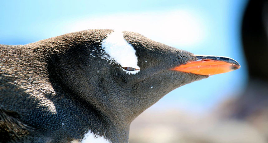
PORTLAND, Ore. — Penguins preserve records of Antarctic environmental change. The birds’ feathers and eggshells contain the chemical fingerprints of variations in diet, food web structure and even climate, researchers reported February 12 at the American Geophysical Union’s 2018 Ocean Sciences Meeting.
The Antarctic environment has changed dramatically in recent decades. Overfishing has led to a decline in krill, small swimming crustaceans that are a key food source for birds, whales, fish and penguins in the Southern Ocean. Climate change is altering wind directions, creating open water regions in the sea ice that become hot spots for life. These changes have cascading effects on food webs and on the cycling of nutrients. “Penguins are excellent bioarchives of this change,” says Kelton McMahon, an oceanic ecogeochemist at the University of Rhode Island in Kingston.
Penguins are at the heart of the Antarctic food web, and their tissues are known to capture details about what they’ve eaten. Different food sources contain different proportions of carbon and nitrogen isotopes, forms of the elements with different numbers of neutrons. For example, food sources such as krill and fish have different amounts of nitrogen-15 relative to nitrogen-14. The tissues of penguins, such as feathers and eggshells, preserve these proportions. Previous studies had already noted a large shift in isotopic values in penguin tissues in the last 80 years, but those studies couldn’t distinguish between shifts in the penguins’ diet versus climate-related shifts in the isotopic values of the microscopic creatures at the very bottom of the food web. So McMahon and his colleagues created a tool to make this distinction — and ultimately to track Antarctic environmental changes through time. The team focused on the isotopic values of individual amino acids, the building blocks of proteins. Those values reveal “a lot about the biochemistry happening inside the body,” McMahon says. Some of these values are significantly altered as food is digested and incorporated into an animal’s body; others are little changed.
To understand what the wild penguins had been eating through time, the team first developed a set of “chemical fingerprints” for the isotopic values of a dozen different amino acids by mapping how the values found in Atlantic herring, a dietary staple, changed in the penguins’ bodies after digestion. The researchers acquired these data through a controlled feeding study in collaboration with the Omaha Henry Doorly Zoo and Aquarium in Nebraska that monitored precisely what, when and how much a population of Gentoo penguins ate. Comparing the chemical fingerprints with wild Gentoo penguin tissues revealed what the wild penguins must have eaten in the past.
Over the past 80 years, the penguins have shifted from eating mostly fish to eating primarily krill and then back to fish, the team reported. There is probably a straightforward historical explanation, McMahon said: In the late 1800s to the mid-1900s, whalers extensively hunted marine mammals that tend to dine on krill. Penguins likely took advantage of the resulting krill surplus. But from the 1970s to 1990s, krill harvesting ramped up, and penguins shifted back to a fish-dominated diet.
But there’s more to the story. Certain amino acids in the penguins’ food are known to pass through the body with their isotopic values essentially unchanged. In fact, the isotopes in those amino acids are thought to reflect the original isotopic values of the creatures at the very base of the Antarctic food web: phytoplankton floating in the Southern Ocean. Because climate drives the isotopic values of those phytoplankton, the isotopes in those amino acids are a record of climate change.
In the case of nitrogen, higher nitrogen-15 values relative to nitrogen-14 in phytoplankton are likely linked to more open water spaces within the sea ice, McMahon said. Such open spaces, called polynyas, have appeared in recent decades around Antarctica due to changes in wind directions linked to climate change.
This study highlights the power of this amino acid isotope technique to track environmental change through animal tissues, says Seth Newsome, an animal ecologist at the University of New Mexico in Albuquerque who was not involved in the study. The technique is becoming popular because it can detect both diet and baseline changes in the food web from the same tissue, he says.
“This 80-year record is just part of a much broader record of change,” McMahon said. The team plans next to look further back in time. Excavations have revealed penguin eggshells as old as 10,000 years that have a story to tell.
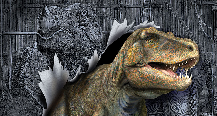
“There’s a very faint dimple here,” Sterling Nesbitt says, holding up a palm-sized fossil to the light. The fossil, a pelvic bone, belonged to a creature called Teleocrater rhadinus. The slender, 2-meter-long reptile ran on all fours and lived 245 million years ago, about 10 million to 15 million years before scientists think dinosaurs first appeared.
Nesbitt, a paleontologist at Virginia Tech in Blacksburg, tilts the bone toward the overhead light, illuminating a small depression in the fossil. The dent, about the size of a thumbprint, marks the place where the leg bone fit into the pelvis. In a true dinosaur, there would be a complete hole there in the hip socket, not just a depression. The dimple is like a waving red flag: Nope, not a dinosaur.
The hole in the hip socket probably helped dinosaurs position their legs underneath their bodies, rather than splayed to the sides like a crocodile’s legs. Until recently, that hole was among a handful of telltale features paleontologists used to identify whether they had their hands on an actual dinosaur specimen.
Another no-fail sign was a particular depression at the top of the skull. Until Teleocrater mucked things up. The creature predated the dinosaurs, yet it had the dinosaur skull depression. The once-lengthy list of “definitely a dinosaur” features had already been dwindling over the past few decades thanks to new discoveries of close dino relatives such as Teleocrater. With an April 2017 report of Teleocrater’s skull depression (SN Online: 4/17/17), yet another feature was knocked off the list.
Today, just one feature is unique to Dinosauria, the great and diverse group of animals that inhabited Earth for about 165 million years, until some combination of cataclysmic asteroid and volcanic eruptions wiped out all dinosaurs except the birds. “I often get asked ‘what defines a dinosaur,’ ” says Randall Irmis, a paleontologist at the Natural History Museum of Utah in Salt Lake City. Ten to 15 years ago, scientists would list perhaps half a dozen features, he says. “The only one to still talk about is having a complete hole in the hip socket.”
The abundance of recent discoveries of dinosauromorphs, a group that includes the dinosaur-like creatures that lived right before and alongside early dinosaurs, does more than call diagnostic features into question. It is shaking up long-standing ideas about the dinosaur family tree.
To Nesbitt, all this upheaval has placed an even more sacred cow on the chopping block: the uniqueness of the dinosaur.
“What is a dinosaur?” Nesbitt says. “It’s essentially arbitrary.” Shared traits In 1841, British paleontologist Sir Richard Owen coined the term “dinosaur.” Owen was contemplating the fossil remains of three giant creatures — a carnivore named Megalosaurus, the plant-eating Iguanodon and the heavily armored Hylaeosaurus. These animals shared several important features with one another but not other animals, he determined. (In particular, he noted, the creatures’ giant legs were upright and tucked beneath their bodies, and each of the animals had five vertebrae fused together and welded to the pelvis.)
Owen decided the animals should be biologically classified together as their own group, or taxon. He named the group “Dinosauria” for “fearfully great lizards.”
In Owen’s day, it was a bit easier to spot similarities between fossils, says paleontologist Stephen Brusatte of the University of Edinburgh. “Back then, there were so few dinosaurs. But the more fossils you find, the patterns become more complicated,” he says. “With every new discovery, you get a different view of what features define a dinosaur. It’s nowhere near as clear-cut as it used to be.”
Dino survivors The largest extinction of species on Earth, the “Great Dying,” happened about 252 million years ago at the end of the Permian Period (SN: 9/19/15, p. 10). About 96 percent of marine species and 70 percent of land species succumbed.
In the period that followed, the Triassic, spanning 252 million to 201 million years ago, new reptilian species arose and flourished. This was the time of the dinosauromorphs, crocodylians (the ancestors of crocodiles) and, of course, the dinosaurs themselves. No one knows exactly when dinosaurs arose, although it was probably around 230 million years ago. For tens of millions of years, the dinosaurs lived alongside numerous other reptile lineages. But at the end of the Triassic, dramatic climate change played a role in another mass extinction. Dinosaurs somehow survived and went on to dominate the planet during the Jurassic Period. Paleontologists once assumed the dinosaurs were somehow superior, with physical features that helped them outcompete the other reptiles. “But that’s not borne out by new dinosaur relatives,” Nesbitt says. Dinosaurs and dinosauromorphs, researchers found, were very similar. The new bonanza of dinosauromorph fossils reveals a repeating pattern of parallel evolution, such as lengthening legs or having legs oriented directly under the body. In short, Nesbitt says, dinosaurs “are not doing anything different than their closest relatives.”
On the heels of those discoveries, many paleontologists suspect that the reason for dinosaurs’ rapid expansion in the Jurassic is simply that the creatures took advantage of the sudden availability of ecological niches left behind by their long-dead cousins from the Triassic.
But that doesn’t explain why dinosaurs survived the extinction at the end of the Triassic, while their dinosauromorph cousins (and most of the crocodylians) died out. That’s a question no one yet has answered.
Maybe dinosaurs had some anatomical characteristics that helped them survive, suggests Max Langer, a paleontologist at the University of São Paulo. “But we don’t know what those features were.” Uprooting the family tree To identify the animal that left behind a fossil, paleontologists pore over the bone, noting each bump, groove and hole, measuring the length of a tibia bone or counting the digits on a forelimb. Before powerful computers were available, scientists constructed evolutionary trees by noting which species share different bumps and grooves, and assessing whether those features (also called characters) were inherited from a common ancestor, or passed along to descendants.
Langer calls that approach to phylogenetic analyses “old-fashioned.” Today, scientists use computer algorithms to help construct elaborate phylogenetic, or evolutionary, trees. But the fossil characters are still the raw data required to create those trees, and the analyses are only as good as those data. Different researchers may choose different features to consider, and may interpret the fossils differently, too. Those concerns hit home among dinosaur researchers last year, when a team proposed a fundamental reorganization of the dinosaur evolutionary tree.
For about 130 years, the basic structure of the dinosaur family tree was considered relatively stable. Dinosaurs were split into two main lines based on the shape of the hips. Both lines had the hole in the hip socket, still considered unique to all dinosaurs. One line known as the ornithischians, also had a pubis bone that pointed down toward the tail. That group includes giant herbivores such as the three-horned Triceratops and plate-armored Stegosaurus. The other line’s pubis bone pointed down toward the front, a hip shape shared by long-necked sauropods such as Brachiosaurus and by carnivorous theropods such as Tyrannosaurus rex. With those hip similarities, sauropods and theropods have long been considered closer “sister” groups, while ornithischians were seen as more distant relations.
But in March 2017, Ph.D. student Matthew Baron and vertebrate paleontologist David Norman of the University of Cambridge, along with paleobiologist Paul Barrett of the Natural History Museum in London, proposed upending that long-standing arrangement.
At the heart of their paper, published in Nature, was the observation that ornithischians have been somewhat overlooked in previous phylogenetic analyses. The herbivorous ornithischians were a really diverse bunch, with a spectacular array of frills and armors and horns and crests.
So the researchers decided to see how different the family tree would look if an analysis included many more ornithischian species. The team incorporated some 457 different fossil characters from 74 species of all kinds of dinosaurs and dinosaur relatives (SN: 4/15/17, p. 7).
The newly constructed tree might as well have been from a whole different forest. It shuffled the three big groups around, putting ornithischians and theropods together into a new group and suggesting that sauropods had split off earlier.
Baron and his coauthors found that the ornithischians had more than 20 features in common with predatory theropods.
The paper made a splash, but many paleontologists were skeptical. The bar to revise a tree that had stood decades of previous phylogenetic analyses ought to be pretty high, Brusatte says.
Indeed, one point arising from the study was just how subjective phylogenetic analyses can be, Irmis says. Which species a study includes clearly affects how the tree turns out, he says. Plus, he adds, “a slight difference in how one person interprets the anatomy of a fossil or a particular character can make a cumulatively huge difference.”
Langer, Brusatte and several paleontologist colleagues decided to tackle the character interpretation part of the problem head on. “When the paper came out, there was this flurry of excitement,” Brusatte says. “But a lot of us noticed right away that there wasn’t a huge amount of description about the characters.” The concern was that, if the fossils weren’t carefully examined and the characters properly assessed, those errors could dramatically skew the results.
So the researchers divvied up the task of traveling around the world to visit the fossils included in the original paper and to reassess all 457 characters described — in person. “It was essentially a replication study,” Brusatte says.
The team went in expecting to cast doubt on the tree created by Baron, Norman and Barrett — or possibly to completely debunk it. But that didn’t exactly happen.
Langer, Brusatte and their coauthors reported last November in Nature that their analyses showed that the original, 130-year-old evolutionary tree was still the best fit to the dinosaur dataset used by Baron’s team.
But, they found, the original tree wasn’t that much more likely to be correct than the newly described tree. “This is the thing that really blew us away: It wasn’t actually a statistically significant result,” Brusatte says. In fact, the often-accepted tree wasn’t even that much more likely than an older, third arrangement of the tree that grouped ornithischians closer to the other herbivores in the family, the long-necked sauropods, and left the fierce theropods as the outliers.
“There is currently great uncertainty about early dinosaur relationships and the basic structure of the dinosaur family tree,” the researchers concluded. “It seems that the flood of new discoveries over the past decades has revealed unexpected complexity.”
Brusatte adds: “We shouldn’t rewrite the textbooks just yet. But we’ve taken what we thought was a certainty and turned it into a mystery — and a big mystery, at that.” Catch-22 How the different dinosaur groups are related to one another may seem like insider baseball, Nesbitt says. But the evolutionary tree is the common ground, the framework within which researchers can discuss dinosaur evolution, dinosaur origins and what binds all dinosaurs together. “It makes it difficult to ask questions about how features are evolving if we can’t have some agreed-upon taxonomy,” he adds.
Similarly, without an agreed-upon evolutionary tree, it’s hard to know which anatomical features to follow through the tree — such as any that might have helped dinosaurs survive the end-Triassic extinction. Each arrangement of the evolutionary tree seems to highlight different features as being particularly important, Langer says. “If you don’t know how the tree is arranged, you can’t say which feature characterizes [dinosaurs].”
The thorny problem revolves around which to tackle first: How to define a dinosaur or how to redraw the dinosaur family tree?
But Langer suggests the answer, as always, is to return to the fossils. In the paper by Langer and his coauthors, they make a plea for researchers to do the mundane work. “We proposed that we need more … anatomical descriptions and definition of characters,” Langer says. “It’s boring to do, but people have to do more of this.”
Finding Teleocrater As Nesbitt cradles the Teleocrater pelvic bone, he turns to a tall cabinet of wide, shallow drawers. He slides open a drawer filled with dozens of carefully labeled boxes, each holding one or more bones from Teleocrater, collected during a 2015 expedition to Tanzania’s Ruhuhu Basin.
The first known fossils of Teleocrater rhadinus, to date the only species of the genus Teleocrater, were actually discovered in the 1930s. But those fossils — a few bits of vertebrae, pelvis and limb — languished unidentified in London’s Natural History Museum for several decades. The Ruhuhu Basin, an area dating to between 247 million and 242 million years ago, was a popular place in the Triassic. The site contains abundant fossils, diverse assemblages of Triassic animals including relatives of crocodylians, giant-headed amphibians and ancient relatives of modern mammals called cynodonts.
In 2010, Nesbitt described a species of dinosauromorph from the Ruhuhu Basin dubbed Asilisaurus kongwe. But on his 2015 expedition, he was hoping to find more evidence that would help identify the mysterious Teleocrater — perhaps even a skull.
He hit pay dirt: His team found a bone bed containing at least three Teleocrater individuals, including a braincase and jawbone. The skull was a particularly exciting find, because it showed the team that Teleocrater, clearly a nondinosaur from other features, had the skull depression, just like a true dinosaur.
Paleontologists tend to say that finding more fossils from early dinosaurs and their close relatives is the surest way to fill in the gaps on how the creatures evolved and to tidy up the family tree.
Nesbitt laughs. “Now we have way more fossils,” he says, “and it’s way messier.”