Chronic flu patients could be an early warning system for future outbreaks

People with weakened immune systems might help scientists get a jump on the flu virus.

Some flu virus mutations popped up again and again in cancer patients with long-term infections, researchers report June 27 in eLife. And some of those mutations were the same as ones found in flu viruses circulating around the world a few years later, evolutionary virologist Jesse Bloom of the Fred Hutchinson Cancer Research Center in Seattle and colleagues discovered. The findings may eventually help vaccine developers predict flu strain evolution.
“You can’t predict what’s going to happen next year,” — at least not yet, Bloom says. But monitoring infections in many people may indicate which parts of the virus are most likely to change in the future.

Most people who catch the flu get over it in about a week. Previous studies have suggested that the virus doesn’t change much within one person. It must pass through tens to hundreds of people to build up enough mutations to give it an advantage over other flu viruses, Bloom says. That makes predicting flu evolution tricky.

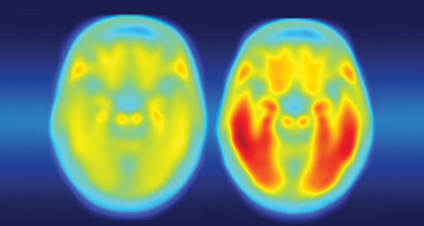
A coffee shop conversation alerted Bloom to a potential treasure: multiple nasal wash samples from four cancer patients who had had the flu for months in 2006 and 2007. Part of their cancer treatments had weakened all four people’s immune systems, making it hard for them to fight off the infections.

Evolutionary biologist Katherine Xue in Bloom’s lab and colleagues examined genetic material from the nasal washes, identifying mutations present in at least 5 percent of flu viruses in each person. The team tracked competition between virus variants within each person over time and compared virus evolution patterns among the patients.

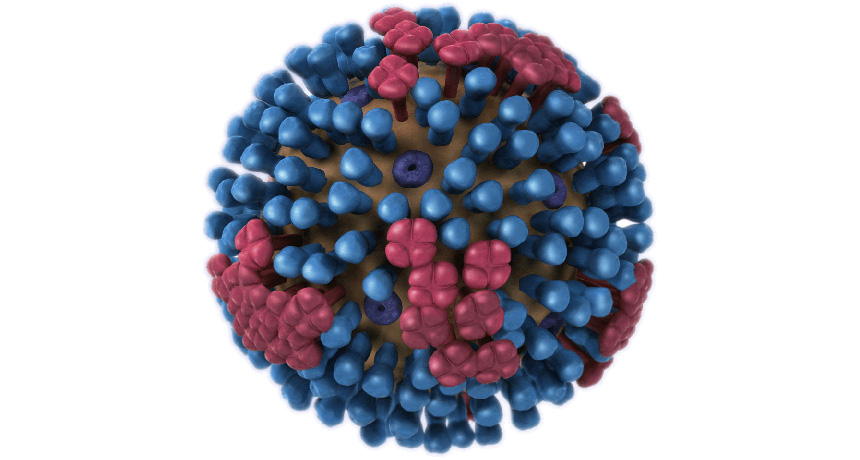
Nine flu mutations popped up in at least two separate patients. Of those, five were in the virus’s hemagglutinin gene. That gene encodes a sugar-studded protein on the virus’s outer surface that helps the virus stick to and invade human cells. The immune system commonly makes antibodies against hemagglutinin that foil the strain’s ability to infect the host again. As a result, the virus has to mutate so that the protein will be different enough to evade the immune system.
Four amino acids of the hemagglutinin protein were frequently changed by mutations in the cancer patients’ viruses and popped up years later in flu strains worldwide, too. Those amino acids are the 138th, 193rd, 223rd and 225th links in the chain of amino acids that make up the hemagglutinin protein.

In some cases, the mutations produced the same amino acid change in both the cancer patients’ and the global virus strains circulating after 2010. For instance, the amino acid valine was altered to isoleucine at position 223. That happened in two cancer patients in 2006 and 2007. After about 2012, nearly all viruses circulating worldwide had the same change.

In another case, those same two cancer patients’ viruses had tyrosine at position 193, but globally circulating viruses had either phenylalanine or serine at that position, the researchers found. Those results indicate that at some spots in the protein, particular changes are important, but other positions are more malleable.

Within patients, viruses carrying different amino acids seemed to directly compete against each other; as one became more frequent, the other was reduced in abundance. That’s the same sort of pattern researchers observe at the global level. Knowing which mutations commonly win competitions in immune-compromised patients may give a preview of winners in the global flu fight.

Not all of the flu mutations that arose in the cancer patients were later found in the general population, says infectious disease biologist Katia Koelle of Duke University. For instance, a mutation called L427F (changing leucine at position 427 to phenylalanine) was found in more than 75 percent of flu viruses in three of the cancer patients, but it was never seen in flu viruses circulating globally. That mutation might give flu viruses some advantage within a person, but might not be efficient at spreading from person-to-person, Koelle says. Studies that compare flu alterations in multiple people won’t immediately tell researchers how to design vaccines, she says, but could point to parts of the virus for further investigation.

Xue and Bloom say they would like to repeat the study, perhaps this time in very young children and elderly people — two groups that also have weaker immune systems than most adults.

Most football players who donated their brains to science had traumatic injury

A majority of football players whose brains were donated for research suffered a degenerative brain disease during their lives, according to the largest sample of players ever studied. The finding provides more evidence that the repetitive injuries to the brain sustained while playing American football are associated with the disease, researchers say.

Of 202 deceased former football players, 177 were diagnosed with chronic traumatic encephalopathy, which can cause a host of mood and behavioral issues as well as thinking and reasoning problems. Among 111 men who had played in the National Football League, 110 — a whopping 99 percent — had developed the disease, researchers report July 25 in JAMA. Three of 14 high school players also showed signs of the brain disease, as well as 48 of 53 college players. Researchers relied on brain autopsies of the players to make the diagnoses and interviewed family members and friends about the symptoms players had experienced.
This doesn’t necessarily mean all football players experience chronic traumatic encephalopathy. Many of the families who donated the brains for research could have been motivated to do so because their loved ones had noticeable symptoms, so the sample is not necessarily representative of the general football population. The results are still worrisome, though, researchers say.
“The fact that chronic traumatic encephalopathy was so common adds to our concern about the safety of playing football and the risk of developing neurologic symptoms later in life,” says neurologist Gil Rabinovici of the University of California, San Francisco, who wrote an editorial accompanying the article. This “hovers like a dark cloud over the game at all levels, even if the study cannot address how frequent the disease is, or who is at risk.”

Chronic traumatic encephalopathy, or CTE, shows up in athletes and others who’ve had repetitive injuries to the head, such as concussions. The only way to diagnose the disease is with an autopsy. In brains with the condition, a protein called tau goes “bad” and forms clumps in nerve cells and other brain cells. Although tau buildup is found in other brain diseases, like Alzheimer’s, in CTE, the protein congregates in brain cells around small blood vessels.
In 2008, a research team set up a brain bank to study the impact of head blows resulting from contact sports or military service. Behavioral neurologist Jesse Mez of Boston University School of Medicine and his colleagues classified players as having mild or severe CTE, depending on how widespread the tau clumps were in the players’ brains. The severity of disease seemed to track with the number of years spent playing football, says Mez. Among NFL players, 95 of the 110 diagnosed cases were severe. All three of the high school players’ cases were mild, while just over half of the college players’ cases were severe.

Yet the players’ reported symptoms while alive were similar, regardless of the severity seen in the brain. Behavioral and mood problems, such as impulsivity, anxiety and depression, were commonly reported in both severe and mild cases of the disease. Cognitive symptoms, including memory loss, were also typical for both groups. One major difference, Mez notes, was that dementia was more common in severe cases of CTE than in mild cases.

As for why players reportedly experienced similar symptoms no matter the severity, “the question is, is there something else going on,” such as inflammation, Mez says. “Or are there regions of the brain that we’re not looking carefully enough at?”

There still isn’t a way to diagnose CTE during life, and that’s “the 800-pound gorilla in the room,” says neurologist David Brody of Washington University School of Medicine in St. Louis.

Detecting the disease in patients will be crucial for understanding how common CTE is in the NFL, “let alone in the millions of people who participated in college, high school and youth football,” says Rabinovici. “In the meantime, we need to focus on prevention of concussions and other head impacts at all levels of contact sports.”

Fossil find suggests this ancient reptile lurked on land, not in the water

A round belly, stubby feet and a tapering tail made one armored reptile a lousy swimmer. Despite earlier reports, Eusaurosphargis dalsassoi might not have swum at all, scientists now say.

E. dalsassoi was first identified in 2003. Fossils were found near Monte San Giorgio at the Swiss-Italian border alongside the remains of marine reptiles and fish that lived roughly 240 million years ago. That association led scientists to conclude the creature was aquatic. But a complete skeleton of E. dalsassoi unearthed in 2002 in the Swiss Alps and recently assembled contradicts that idea.
At just under 20 centimeters long, the fossil, probably of a youngster, shows that E. dalsassoi widened at the stomach and slithered forward with stiff elbow and knee joints and spadelike claws. That’s not a swimmer’s build, paleontologist Torsten Scheyer of the University of Zurich and colleagues report June 30 in Scientific Reports.

Armed with rows of small spikes along its back and spear-shaped plates framing its head, sides and tail, the animal resembled today’s girdled lizards. The researchers speculate that this particular E. dalsassoi died on a beach and then got washed into the ocean.

How an itch hitches a ride to the brain

Scientists have traced the sensation of itch to a place you can’t scratch.

The discomfort of a mosquito bite or an allergic reaction activates itch-sensitive nerve cells in the spinal cord. Those neurons talk to a structure near the base of the brain called the parabrachial nucleus, researchers report in the Aug. 18 Science. It’s a region that’s known to receive information about other sensations, such as pain and taste.

The discovery gets researchers one step closer to finding out where itch signals ultimately end up. “The parabrachial nucleus is just the first relay center for [itch signals] going into the brain,” says study coauthor Yan-Gang Sun, a neuroscientist at the Chinese Academy of Sciences in Shanghai.
Understanding the way these signals are processed by the brain could someday provide relief for people with chronic itch, Sun says. While the temporary itchiness of a bug bite is annoying, longer term, “uncontrollable scratching behavior can cause serious skin damage.”

Previous studies have looked at the way an itch registers on the skin or how neurons convey those sensations to the spinal cord. But how those signals travel to the brain has been a trickier question, and this research is a “major step” toward answering it, says Zhou-Feng Chen, director of the Center for the Study of Itch at Washington University School of Medicine in St. Louis.

A network of neurons in the spinal cord wrangles itch signals, previous research suggests. In particular, spinal neurons that make a protein called gastrin-releasing peptide receptor have been shown to be important in itch signaling. But those neurons didn’t link up directly to the parabrachial nucleus, or PBN, Sun’s team found; instead, they talked to other neurons that send messages to the PBN.

When mice were given injections of a drug that induces allergic itching, the rodents showed greater activity in those neurons connecting the spinal cord to the PBN, Sun and colleagues found. In another experiment, the researchers made neurons going to the PBN light-sensitive, and then used light to stop those neurons from sending messages. When those nerve cells were blocked, mice given an itch-triggering drug scratched less.

It’s too soon to say whether itch signals in humans follow the same route — or whether all kinds of itches take the same path. An allergic itch is different from the sort of itch that comes from a light touch, and the two might be handled differently by the brain (SN: 11/22/08, p. 16). And mice, unlike humans, can’t actually describe how itchy they’re feeling. So scientists have to rely on clues like scratching, a reaction to an itch, not a direct measurement of the sensation itself. Which raises the question: If you don’t feel an urge to scratch an itch, is the itch really there at all?

Gene variant linked to Alzheimer’s disease is a triple threat

A genetic risk factor for Alzheimer’s disease is a double, make that triple, whammy.

In addition to speeding up the development of brain plaques associated with Alzheimer’s, a gene variant known as APOE4 also makes tau tangles — another signature of the disease — worse, researchers report online September 20 in Nature. APOE4 protein also ramps up brain inflammation that kills brain cells, neuroscientist David Holtzman of Washington University School of Medicine in St. Louis and colleagues have discovered.
“This paper is a tour de force,” says Robert Vassar, a neuroscientist at Northwestern University Feinberg School of Medicine in Chicago. “It’s a seminal study that’s going to be a landmark in the field” of Alzheimer’s research, Vassar predicts.

For more than 20 years, researchers have known that people who carry the E4 version of the APOE gene are at increased risk of developing Alzheimer’s. A version of the gene called APOE3 has no effect on Alzheimer’s risk, whereas the APOE2 version protects against the disease. Molecular details for how APOE protein, which helps clear cholesterol from the body, affects brain cells are not understood.

But Holtzman and other researchers previously demonstrated that plaques of amyloid-beta protein build up faster in the brains of APOE4 carriers (SN: 7/30/11, p. 9). Having A-beta plaques isn’t enough to cause the disease, Holtzman says. Tangles of another protein called tau are also required. Once tau tangles accumulate, brain cells begin to die and people develop dementia. In a series of new experiments, Holtzman and colleagues now show, for the first time, that there’s also a link between APOE4 and tau tangles.
In one experiment, mice that had no A-beta in their brains developed more tau tangles if they carried the human version of APOE4 than if they had the human APOE3 gene, Holtzman and colleagues found. That finding indicates APOE4 affects tau independently of A-beta.
Brains of people who died from various diseases caused by tangled tau had more dead and damaged cells if the people carried APOE4. The researchers also tracked 592 people who had low levels of A-beta in their cerebral spinal fluid — a clue that plaques have formed in the brain — and who showed symptoms of Alzheimer’s. Over a five- to 10-year period, the disease progressed 14 percent faster in people with one copy of APOE4 and 23 percent faster in people with two copies than in people who didn’t have that version of the gene, the researchers found. Those worsening symptoms are presumed to be caused by more rapid buildup of tau tangles in the APOE4 carriers.

APOE4 also seems to make Alzheimer’s worse by causing inflammation, the researchers found. Two kinds of mouse glial brain cells, microglia and astrocytes, making different versions of the APOE protein were grown with brain nerve cells, or neurons, that make disease-causing forms of tau. Mouse neurons grown with glia making no APOE grew well, even though they were making abnormal tau. But neurons grown with glia making APOE4 often died. APOE4 provoked inflammation responses in the normally friendly astrocytes and microglia, leading those cells to kill neurons, the researchers found. Such inflammation can make brain degeneration worse.

The data linking the APOE4 gene to tau tangles and brain inflammation is “super tight,” says molecular neurobiologist Sangram Sisodia of the University of Chicago. But the molecular details behind how APOE4 protein causes those effects are still vexingly absent, he says. Much more work is needed to uncover which molecules APOE4 interacts with, so that researchers can devise ways to counteract its negative effects in the brain.

Any therapies that decrease or eliminate APOE4 will need to be limited to the brain, because the protein is needed in the rest of the body to maintain healthy cholesterol levels, Vassar says. “You don’t want to give a person heart disease to cure Alzheimer’s disease.”

How a meteor shower helped solve the case of the vanishing comet

The reappearance of a long-lost meteor shower has finally explained what happened to a missing comet named 289P/Blanpain.

That comet was spotted only once in 1819 and never again, unusual for a body orbiting the sun. But in 2003, astronomers found a small asteroid moving along the Blanpain orbit, suggesting the space rock might be the comet (or a piece of it) after it ejected much of its cometary dust.

Some of that dust may have been what Japanese researchers saw in 1956 when they observed a meteor shower from the constellation Phoenix. Meteor showers occur when dust left behind by a comet burns up as it hits Earth’s atmosphere. Those “Phoenicid” meteors hadn’t been seen before — or since.
Astronomer Jun-ichi Watanabe of the National Astronomical Observatory of Japan in Tokyo and colleagues traced the meteors to where the comet’s dust trail should have been. In 2010, the group predicted that the remaining dust would create another shower in 2014.

Team members traveled to North Carolina and Spain’s Canary Islands to test their prediction, and on the first two days of December, 2014, they saw Phoenicids streak across the sky. But there were about 90 percent fewer meteors than expected; Blanpain may have lost its dust more quickly than previously thought, the team reports in the Sept. 1 Planetary and Space Science. The astronomers will get a second chance to check — another shower is expected in 2019.

When it comes to self-driving cars, what’s safe enough?

Self-driving vehicles passed a major milestone in November when Waymo’s minivans hit the streets of Phoenix without backup human drivers — reportedly making them the first fleet of fully autonomous cars on public roadways. Over the next few months, people will get a chance to take these streetwise vehicles for a free spin as the company tries to drum up excitement — and a customer base — for its launch of a driverless taxi service.

But even as these cars are ditching human supervisors, many people doubt the safety of machine motorists. A whopping 85 percent of baby boomers and even 73 percent of millennials confess to being afraid to ride in self-driving cars, according to a recent AAA survey. And while Waymo claims its vehicles are designed to be the world’s most experienced drivers — based on road tests as well as clocking millions of virtual miles — there’s still no consensus among experts about how safe is “safe enough” when it comes to street-smart cars.
It’s especially difficult to tell whether self-driving cars have earned their licenses when scientists are still writing the driver’s test.

Besides the sheer convenience of being able to take your hands off the wheel, the major appeal of self-driving cars is safer roadways. After all, mechanical chauffeurs can’t get drunk or distracted — factors involved in 29 and 10 percent of fatal accidents, respectively. But the only surefire way to evaluate autonomous cars’ reliability is test-driving them in real traffic, explains Nidhi Kalra, an information scientist at the RAND Corporation in San Francisco. “I think a lot of people were thinking, ‘Oh, we’ll just wait until the companies do enough test-driving,’” she says. “You could wait until the next millennium until that happens.”

In a 2016 study, Kalra and a colleague showed that self-driving cars would have to trek hundreds of millions or perhaps billions of miles to demonstrate with comfortable certainty that they caused fewer fatalities than the average person (about 1.1 per 100 million miles driven). Based on the current number of self-driving cars, that task could take decades or centuries to complete.

Tech developers hardly have that kind of time, so companies like Waymo assess their vehicles’ safety by pairing real driving time with practice on a private track and millions of miles a day in computer simulations.
Still, simulations can’t replace the value of actual road experience, says Philip Koopman, an electrical and computer engineer at Carnegie Mellon University in Pittsburgh. “What about the scenarios they didn’t know [to simulate]?” he says. “Weird, weird, weird stuff happens out on the roadways.”

Since current self-driving safety assurances aren’t exactly airtight, Koopman argues that self-driving cars should be held to a way higher standard than human drivers — say, 10 times safer than the average human — before they’re given the green light. That would provide enough wiggle room in the margin of error to assume that the driverless car actually is safer, Koopman reasons.

But getting to that point could take a long time, and miss the opportunity to save many lives, Kalra says. She’s confident because her team forecast a future — actually lots of different futures — where self-driving cars hit the road when they were 10, 75 or 90 percent safer than the average human driver. At 10 percent, fatalities drop to one death per 100 million miles. Maybe that doesn’t seem like a lot, but with those cars much closer to being ready to roll, some 500,000 lives could be saved between 2020 and 2050, the team forecasts, compared with the imagined futures where people hold out for way higher safety standards.

But just aiming for 10 percent safer doesn’t provide much margin for error, Koopman argues. “You’re cutting it pretty close.”

And a lower safety standard could mean more accidents at first — and a public backlash, says Azim Shariff, a psychologist at the University of California, Irvine. People may be less inclined to accept mistakes made by machines than humans, and research has shown that people are more risk-averse when it comes to risks that they can’t control.

“What happens when a 4-year-old in the back of a car that’s operated by her mother gets killed by an autonomous car?” Shariff asks.

Success depends on buy-in. “So public opinion is really going to matter,” Shariff says.

Right now, most Americans may not be lining up to hop aboard fully autonomous cars. But “once people start knowing people who have been in them and lived to tell the tale, so to speak, I think it will change quickly,” says David Groves, a policy analyst at the RAND Corporation in Santa Monica, Calif.

Kalra also suspects that people will fear autonomous cars less when the National Highway Traffic Safety Administration establishes a self-driving car safety rating, like its crash test ratings for traditional cars. That kind of rating system “will probably come after the technology is on the road, just as it did for regular cars,” she says. “We didn’t have a safety rating system when the Model T came out. It sounds like it’s the cart before the horse to have cars before safety ratings, but that’s often how it happens.”

‘Death: A Graveside Companion’ offers an outlet for your morbid curiosity

Death: A Graveside Companion makes for an unusual coffee-table book, with its coppery etched Grim Reaper on the cover. Yet you may be surprised by how much fun it is to pore through the book’s lavish artwork of skulls, cadavers and fanciful imaginings of the afterlife.

There is, after all, a reason for the term “morbid curiosity.” It’s only natural for people to try to understand and come to terms with their inevitable demise, and as the book reveals, it is only in modern Western society that the topic of death has become so taboo. Even as recently as Victorian times, the book notes, the dead were laid out in the family parlor, their hair cut off and twisted to make decorative mementos to hang on the wall.
As a founder of New York City’s now-closed Morbid Anatomy Museum, Joanna Ebenstein has set out to help change modern attitudes, by giving us permission to let our morbid curiosity loose. “It is my hope that this book might act as a gesture towards redeeming death, to invite it back into our world in some small way,” she writes. “It is precisely by keeping death close at hand and coming to terms with its inevitability that we are able to lead full rich lives.”
She brings together 1,000 images of historical artwork, illustrations and artifacts showcasing humankind’s ongoing quest to imagine and find meaning in death, along with 19 essays by a diverse set of writers, art experts and scientific thinkers. The writings cover spiritual and symbolic aspects of death, such as the origins of Mexico’s Day of the Dead, and the surprising variety of death-themed amusements over the years. An early Coney Island attraction, for instance, re-created the experience of being buried alive. Some essays delve into scientific history, such as miniature crime scenes used in forensic science and the history of cadavers in the study of anatomy.
While the essays are illuminating, the illustrations and photographs, along with informative captions, provide most of the book’s substantial heft, as well as its heart. Only by browsing through still life paintings called vanitas, popular in the 16th and 17th centuries, for instance, will you truly grasp what these symbolic masterpieces are meant to convey: the transience of beauty and earthly pursuits.
If I have any quibble with this compendium, it’s that the essays (but thankfully not captions) are printed in sepia tones that make them hard to read without good lighting. But given the subject, this book may be best read while sitting next to a sunny window anyway.